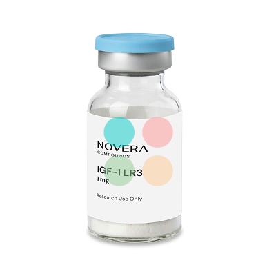
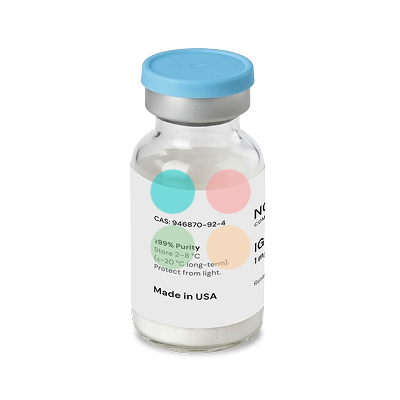
IGF-1 LR3 is a synthetic analog of human insulin-like growth factor-1 (IGF-1), engineered with two structural modifications that reduce its affinity for IGF binding proteins and extend its biological activity in preclinical research models. Researchers studying cell proliferation, metabolic signaling pathways, and tissue repair use this 83-amino-acid peptide because its prolonged half-life of 20 to 30 hours allows sustained receptor activation in both cell culture and animal models. IGF-1 LR3 is not FDA approved for human therapeutic use, and all evidence discussed below comes from in vitro or animal studies.
FillerSupplies.com supplies receptor grade IGF-1 LR3 in lyophilized powder form for laboratory research use only. Licensed researchers and medical professionals seeking to buy IGF-1 LR3 online can rely on the Novera brand for consistent compound quality, temperature-controlled shipping, and expert technical support.
General Information About IGF-1 LR3
Native human IGF-1 is a 70-amino-acid polypeptide that signals through the IGF-1 receptor (IGF-1R), a receptor tyrosine kinase on the cell surface. IGF-1 LR3 retains the full core sequence but carries two structural modifications: an arginine substitution replacing glutamic acid at position 3, and a 13-amino-acid extension at the N-terminus (sequence MFPAMPLSSLFVN). These changes yield an 83-amino-acid molecule with a molecular weight of approximately 9,111 Da and a molecular formula of C₄₀₀H₆₂₅N₁₁₁O₁₁₅S₉.
The arginine substitution and N-terminal extension sit outside the principal receptor-binding domains, so IGF-1 LR3 retains full affinity for the IGF-1 receptor. Upon binding IGF-1R, the peptide activates the PI3K/Akt and MAPK/ERK signaling cascades, which regulate protein synthesis, cell survival, and metabolic function. IGF-1 LR3 activates the PI3K signaling pathway with the same downstream consequences as native IGF-1, but the free fraction persists longer in circulation.
What distinguishes the compound from native IGF-1 is its interaction profile with IGF binding proteins (IGFBPs). Francis et al. reported in 1992 that LR3-IGF-1 exhibits roughly 1,000-fold lower affinity for IGFBP-3 and IGFBP-4 compared to native IGF-1. Because native free IGF-1 has a plasma half-life of only 10 to 15 minutes when unbound, while IGF-1 LR3 has an extended half-life of 20 to 30 hours in animal models, the analog provides continuous cellular signaling over a period that would require repeated dosing with the native peptide. IGF-1 LR3 maintains continuous cellular signaling due to its prolonged activity, making it useful for studies requiring sustained IGF-1R stimulation in controlled laboratory settings.
FillerSupplies.com provides this compound as receptor grade lyophilized powder for laboratory research use, suitable for reconstitution in sterile water or appropriate buffer systems.
IGF-1 LR3 Use in the Research Setting
Supplied as a lyophilized powder intended for research and laboratory use only, IGF-1 LR3 is primarily used in research settings spanning cellular proliferation, metabolic signaling, skeletal muscle therapeutics, and regenerative medicine. The subsections below summarize the current preclinical evidence base.
Cellular Proliferation and Growth Studies
IGF-1 LR3 promotes cellular proliferation across multiple tissue types. In a 1992 study by Francis and colleagues published in the Journal of Molecular Endocrinology, LR3-IGF-1 demonstrated 5- to 10-fold greater biological potency than native IGF-1 in promoting cell proliferation of cultured L6 rat myoblasts. The enhanced mitogenic activity stems from the peptide’s reduced sequestration by IGF binding proteins, leaving a larger free fraction available to bind the IGF-1 receptor on the cell surface.
Downstream of IGF-1R binding, the compound activates PI3K/Akt signaling (promoting cell survival and protein synthesis) and the MAPK/ERK cascade (driving cell division). IGF-1 LR3 may stimulate cell division in muscle and bone tissue, and it enhances anabolic activities by 2.5-fold over native IGF-1 in comparable experimental systems.
“In vitro L6 myoblasts showed LR3-IGF-1 achieved 5- to 10-fold higher proliferation than native IGF-1 due to poor IGFBP binding.” (Francis et al., 1992, Journal of Molecular Endocrinology)
In a 14-day whole-animal study (female rats), Francis et al. found that LR3 was approximately 6-fold more potent than native IGF-1 in measures of body weight gain, nitrogen retention, and food conversion efficiency. IGF-1 LR3 provides almost three times cellular activation compared to IGF-1 in certain proliferation assays, and IGF-1 LR3 exposure may enhance cellular turnover in livestock models where systemic administration is feasible.
Citation capsule: Francis et al. (1992) demonstrated in female rats that 14-day systemic administration of LR3-IGF-1 produced 6-fold greater growth promotion than equimolar native IGF-1, measured by nitrogen retention and body weight gain, attributable to the analog’s 1,000-fold lower IGFBP-3 binding affinity.
Metabolic Signaling Research
IGF-1 LR3 enhances GLUT4 translocation to the cell surface in muscle cells and adipocytes, a mechanism mediated by PI3K/Akt activation and downstream phosphorylation of AS160/TBC1D4. Through this pathway, IGF-1 LR3 may improve glucose uptake in muscle cells and may reduce blood sugar levels in animal models. Hypoglycemia can occur due to IGF-1 LR3 mimicking insulin activity, a finding confirmed in multiple species.
Because the analog stays free in circulation rather than bound to IGFBPs, animal work has repeatedly recorded stronger and longer glucose-lowering than an equivalent dose of native IGF-1. That behaviour underscores the compound’s relevance to metabolic research exploring glucose transporters, insulin levels, and insulin receptor cross-reactivity.
IGF-1 LR3 may alter AMPK activity in cells, adding another layer to its metabolic signaling profile. Studies in mice also focus on IGF-1 LR3’s effects on aging, while separate preclinical work suggests the compound may influence kidney disease progression in research models.
Muscle Preservation and Myostatin Interaction
Myostatin (growth differentiation factor 8) suppresses muscle cell growth and differentiation by activating the Smad3 signaling axis. Research suggests that IGF-1 LR3 may counteract myostatin’s inhibitory effects through the PI3K/Akt pathway, which blocks Smad3 activation and promotes expression of the myogenic transcription factor MyoD. MyoD activation by IGF-1 LR3 may promote muscle growth by driving terminal differentiation of myoblasts.
IGF-1 signalling through PI3K/Akt drives protein synthesis and blocks the atrophy programme myostatin promotes, a balance Yoshida and Delafontaine set out in their 2020 Cells review of skeletal-muscle hypertrophy and atrophy. The same mechanism is presumed for LR3, which binds IGF-1R with comparable affinity.
IGF-1 LR3 enhances muscle cell proliferation in experimental models and reduces muscle protein breakdown by up to threefold compared to untreated controls. Research indicates IGF-1 LR3 may mitigate muscle atrophy progression, and the peptide stimulates systemic and local protein synthesis, making it a tool for studying skeletal muscle therapeutics and myostatin attenuation in preclinical research models.
<em>Important to Know:</em>
Comparative IGF Analog Studies
Researchers working with multiple variants of insulin-like growth factor often need to select between native IGF-1, IGF-1 LR3, and DES(1-3)IGF-1 based on experimental requirements. The table below summarizes the key differences relevant to study design:
|
Characteristic
|
Native IGF-1
|
IGF-1 LR3
|
DES(1-3)IGF-1
|
|
Amino Acid Length
|
70 aa
|
83 aa (70 core + 13-aa N-terminal extension)
|
67 aa (lacks first 3 aa of native sequence)
|
|
IGFBP Binding Affinity (relative to native)
|
High; baseline
|
~100- to 1,000-fold reduced
|
~10-fold reduced
|
|
Half-Life (Free / Systemic)
|
10-15 min free; 12-16 h bound in ternary complex
|
~20-30 hours in animal models
|
~20-30 min
|
|
IGF-1R Affinity
|
Baseline (1×)
|
Equivalent to native; enhanced receptor availability due to reduced IGFBP buffering
|
Slightly elevated free-fraction effect
|
|
Insulin Receptor Cross-Reactivity
|
Low at physiological levels
|
Low to moderate (model-dependent)
|
Higher, particularly in fasted models
|
|
Primary Research Applications
|
Physiological regulation; cartilage, bone, metabolic disease
|
Muscle growth, systemic metabolic signaling, long-exposure studies
|
Local tissue repair, short-acting receptor signaling, wound sites
|
DES(1-3)IGF-1 lacks the N-terminal Gly-Pro-Glu tripeptide, which reduces but does not eliminate IGFBP binding. Its half-life of roughly 20 to 30 minutes makes it suited for localized applications, while IGF-1 LR3’s 20- to 30-hour half-life favors systemic or sustained-exposure protocols. IGF-1 LR3 may enhance cellular activation nearly threefold compared to native IGF-1 when measured across proliferation endpoints, partly because its reduced IGFBP interaction profile leaves more free peptide available for receptor binding.
Tissue Repair and Regeneration Research
IGF-1 LR3 may promote tissue repair and cell survival through sustained IGF-1R activation and downstream protein synthesis. The compound accelerates the healing of connective tissues and ligaments in animal models, a property investigated across tendon, cartilage, and wound-healing research.
In a rat model of tendon transection and overload, Kurtz et al. found that local injection of 25 µg LR3-IGF-1 in methylcellulose gel reduced functional recovery time: animals reached 50% recovery by day 9 compared to day 13 in untreated controls. Collagen content (measured by hydroxyproline assay) and DNA content both increased in treated tendons, suggesting the peptide enhanced both matrix deposition and cellular proliferation at the repair site.
Cartilage repair research using native IGF-1 in rabbits with full-thickness articular defects showed improved neo-cartilage formation by weeks 4 through 8 at higher doses. Separately, a one-year minipig study demonstrated that sustained IGF-1 overexpression via rAAV vector and alginate hydrogel improved cartilage histology, cell density, and collagen deposition while reducing osteoarthritis progression and inflammation. These findings, while obtained with native IGF-1 rather than LR3 specifically, support the broader role of insulin-like growth factor receptor signaling in regenerative medicine.
Citation capsule: Kurtz et al. reported in a rat tendon transection model that 25 µg of locally injected LR3-IGF-1 accelerated 50% functional recovery from day 13 (control) to day 9 (treated), with concurrent increases in hydroxyproline and DNA content at the repair site.
<em>Important to Know:</em>
Buy IGF-1 LR3 Online at FillerSupplies.com
FillerSupplies.com is the best place to buy IGF-1 LR3 for laboratory research. The Novera-brand receptor grade IGF-1 LR3 is supplied as lyophilized powder, ready for reconstitution in your preferred buffer system. Whether your work involves cellular proliferation assays, metabolic research, or tissue repair protocols, you can buy receptor grade IGF-1 LR3 with confidence from an established supplier serving the research community since 2006. IGF-1 LR3 should only be purchased as a research chemical, and many online sources selling IGF-1 LR3 may lack quality controls; FillerSupplies.com addresses this concern through the following commitments:
-
Authentic, certified products: original, quality-controlled compounds, warehouses worldwide.
-
11 years of reliability & trust: established supplier on the market since 2006.
-
Temperature-controlled shipping: thermal packaging with ice gel / cold packs preserves stability, critical for peptides sensitive to repeated freeze thaw cycles.
-
Express worldwide shipping: next-day dispatch, global delivery.
-
Best wholesale prices: tiered bulk discounts and price-match guarantee.
-
Privacy guaranteed; expert support at (888) 392-6640, WhatsApp and email, Mon-Fri 10am-6pm EST.
-
Supplied for research use only, to licensed professionals.
Purity of research peptides is typically assessed by high-performance liquid chromatography. All products from FillerSupplies.com are intended for laboratory research use only and are sold exclusively to licensed professionals.
Related research peptides available at FillerSupplies.com: Follistatin 344, Hexarelin, GHRP-6.
FAQ
What Is IGF-1 LR3 And How Does It Work?
IGF-1 LR3 is a synthetic analog of human insulin-like growth factor-1, modified with an arginine substitution at position 3 and a 13-amino-acid extension at the N-terminus. It binds the IGF-1 receptor (a receptor tyrosine kinase) and activates the PI3K/Akt and MAPK/ERK pathways, promoting protein synthesis, cell proliferation, and cell survival. All current evidence for its biological activity comes from in vitro and animal studies.
What Is The Difference Between IGF-1 LR3 And Native IGF-1?
Native IGF-1 contains 70 amino acids and binds tightly to IGF binding proteins, giving it a free half-life of 10 to 15 minutes. IGF-1 LR3 contains 83 amino acids with roughly 1,000-fold lower IGFBP affinity, resulting in a half-life of 20 to 30 hours in animal models. Francis et al. (1992) found LR3 was approximately 6-fold more potent than native IGF-1 in rat growth assays.
How Does IGF-1 LR3 Compare With DES(1-3)IGF-1?
DES(1-3)IGF-1 is a truncated 67-amino-acid form with a half-life of only 20 to 30 minutes, suited for localized tissue repair studies. IGF-1 LR3 has a 20- to 30-hour half-life and roughly 100- to 1,000-fold lower IGFBP binding, making it more appropriate for systemic or sustained-exposure experimental models. The two analogs serve complementary roles depending on whether local or systemic receptor signaling is the research target.
What Are Typical Research Dosing Ranges For Cell Culture And Animal Studies?
For local tissue repair, Kurtz et al. injected 25 µg of LR3-IGF-1 in methylcellulose gel in a rat tendon model. Systemic dosing in animal models varies by species and study design; researchers should consult the primary literature for their specific model organism.
How Should IGF-1 LR3 Be Stored And Reconstituted?
Lyophilized powder should be stored at −20 °C. Reconstitute with sterile water or an appropriate buffer (such as 0.1 M acetic acid with 0.1% BSA), aliquot into single-use volumes, and store reconstituted aliquots at −20 °C. Avoid repeated freeze thaw cycles, as these degrade peptide integrity and disulfide bond stability.
What Safety Concerns Exist In Research Settings?
Hypoglycemia can occur due to IGF-1 LR3 mimicking insulin activity, particularly at higher systemic doses in animal models. The peptide's mitogenic properties mean IGF-1 LR3 could theoretically accelerate tumor growth in models containing transformed cells. Studies in mice focus on IGF-1 LR3's effects on aging, and the compound may influence kidney disease progression in research; careful dose selection and monitoring are essential.
Is IGF-1 LR3 Prohibited In Competitive Sports?
IGF-1 LR3 is banned by the World Anti-Doping Agency under the S2 category (peptide hormones, growth factors, and related substances). Any use of IGF-1 analogs that activate the IGF-1 receptor is prohibited for performance enhancement. Research applications must be clearly separated from any competitive sport context.